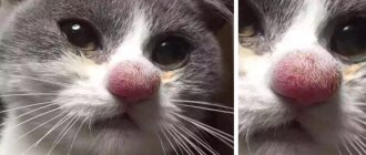
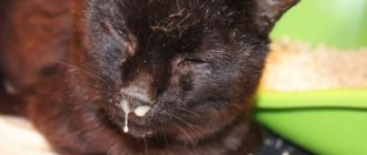
If black dots appear on a cat’s nose, owners should not immediately worry. Such spots may be ordinary moles that people also have. But still, when a kitten has dark or white crusts around its mouth, it is recommended to show your pet to a veterinarian. Often the problem is associated with a skin disease and requires urgent treatment. At home, it is impossible to determine why a cat’s nose darkens, but a veterinary clinic will help determine the cause through a series of diagnostic examinations.
Lentigo - cat freckles
Lentigo is a genetic disorder that causes dark, freckle-like spots to appear. The spots are either black or brown. When you feel the spots, you will find that they are flat or slightly convex. Freckles have clearly defined edges and a diameter of 1–10 mm. The skin around the spots is of normal color.
If your cat only has a few freckles, the condition is called lentigo simplex. If there are a lot of freckles and they form spots of a large area, this is called – which gather so many that they merge into large spots of large freckles, this is called lentiginosis.
Freckles are the result of pigment cells called melanocytes. They contain more melanin than the surrounding skin. No one knows why some cats have a genetic predisposition to lentigo, while other cats of the same color are not prone to freckles.
Calcivirosis
Calcivirosis is a dangerous viral disease of cats. In addition to the nose and oral cavity, the infection can affect the respiratory, hematopoietic and nervous systems of animals, and the digestive organs. The disease is often severe, with symptoms appearing within 1-3 weeks, and about a third of those affected die. Survivors are able to shed the virus into the environment for up to 12 months. With stress, deterioration of living conditions, infection with VLK or FIV, a relapse of calicivirus occurs. If a secondary infection occurs, the mortality rate increases to 80%.
To avoid death, you need to show the animal to a veterinarian as soon as possible.
Lentigo - a feature of saffron milk caps
Since lentigo is a genetic disease, it is associated with color genes. There is no evidence, but observations show that freckles are more likely to form in cats and cats with a red color. Moreover, freckles begin to form in adulthood and old age.
Cats of red, red, tortoiseshell and fawn colors are prone to lentigo. Obviously, all these varieties have the same color gene. Cream and silver cats also sometimes have freckles, but this is the exception to the rule.
The first lentigines usually form on the lips and gums, and then on the nose. The process begins when the cat is 1–3 years old. As your cat ages, freckles appear on other parts of the body and increase in number and size. After appearing on the nose, the spots “migrate” to the area of the ears and paw pads.
Treatment of fungus on a cat's nose
A fungus on the cat’s nose was treated with a course of injections. At the first appointment, the doctor shone ultraviolet light on the spot and confirmed that it was a fungus. The patient had to be given an injection.
Course duration and injection frequency:
- 07/27/2020 - 1st antifungal injection (photo at the beginning of the article was taken on 07/26/2020).
- 08/06/2020 — 2nd injection.
- 08/16/2020 - 3rd injection.
The article was written much later than these dates, but the numbers were written down in the cat’s veterinary passport. Photos were taken periodically and saved on the phone.
After the first injection, the cat returned from the clinic with a significantly lighter nose. However, after some time, the stain began to appear again. And this happened every time: after the injection, the nose became lighter, and a little later it darkened. The injections were aimed at developing immunity to the fungus, and as the body fought the fungus, the spot either faded or darkened.
The cat was prohibited from swimming during treatment. However, he didn't mind :)
On August 18 (a couple of days after the last injection) the cat looked like this:
However, the spot later reappeared (albeit to a lesser extent). The veterinarian recommended coming for an examination two weeks after the course, so the poor fellow was brought to the clinic only a couple of weeks later. The doctor shone an ultraviolet light and said that the situation is much better, the fungus is almost not visible: we need to wait a couple more weeks.
Indeed, the stain soon disappeared. During the treatment, the cat behaved normally: ate, played, etc. Unless I traditionally threw tantrums when going to the clinic :)
So antifungal therapy is a long-term thing. And the problem needs to be solved with the help of a doctor.
This is what this handsome guy looks like now:
The nose is completely clean - the fungus has been completely defeated!
Sunlight has no effect on lentigo
It is known that in humans, freckles become more intense in color in the spring and summer. Lentigines are not exactly freckles (although they are similar in “behavior” and appearance) and they do not depend on sunlight.
However, direct sunlight must be treated with care. Lentigines may darken slightly due to tanning, and the skin around them may become damaged due to burns. Since the nature of lentigines is still not understood, many veterinarians assume that the bulging growths can develop into melanoma (skin cancer).
Important! It is lentigo that often causes the first symptoms of skin cancer – dark, raised spots – to be missed.
Eye diseases in humans: list, symptoms
The reason for this is many factors. For example, the rapid development of computer technology and the deterioration of the environmental situation every year. Next, we will consider the most common diseases, and also highlight their characteristic symptoms.
Pathology of the optic nerve
Glaucoma
- a chronic disease. Due to increased pressure inside the eyes, optic nerve dysfunction occurs. As a result, vision decreases, which may disappear in the future. The disease progresses very quickly, so the patient risks completely losing his vision if he delays going to the doctor. Signs: impaired lateral vision, black spots, “hazy” images, inability to distinguish objects in the dark, colored rings appear in bright light.
Ischemic optic neuropathy
– circulatory disorders in the intraocular or intraorbital region. Symptoms: decreased visual acuity, appearance of “blind” spots in some areas. Reducing viewing angle.
Ischemic neuropathy
Neuritis
- infection. An inflammatory process in the optic nerve is characteristic. Signs: loss of sensitivity in the area around the eye, pain, weakening of the muscles associated with the optic nerve.
Nerve atrophy
– a disease characterized by dysfunction of arousal conduction. Color perception and viewing angle are impaired. Vision decreases and a person can become completely blind.
Nerve atrophy
Pathology of the eye orbit, eyelids, lacrimal canals
Blepharitis
- inflammation that occurs along the edges of the eyelids. Symptoms: swelling of the tissue, accompanied by burning and redness. The patient feels as if a speck has gotten into his or her eye. There is itching and characteristic discharge. Bright light is difficult to perceive, tearing, pain. Dry eyes and peeling of the eyelid margins may occur. After sleep, purulent scabs form on the eyelashes.
Blepharitis
Cryptophthalmos
- a common disease in which the edges of the eyelids fuse together. This causes the palpebral fissure to narrow or even disappear.
Lagophthalmos
– a pathology characterized by a violation of the closure of the upper and lower eyelids. As a result, some areas remain open all the time, including during bedtime.
Turn of the century
– the place where eyelashes grow is turned towards the eye socket. This creates severe discomfort due to rubbing and irritation of the eyeball. Small ulcers may form on the cornea.
Turn of the century
Coloboma of the century
- disturbance in the structure of the eyelids. Usually occurs along with other morphological defects. For example, cleft palate or cleft lip.
Swelling of the eyelid
– localized accumulation of excess fluid in the tissues around the eyelid. Symptoms: local redness of the skin, discomfort. Eye pain worsens when touched.
Swelling of the eyelid
Blepharospasm
- looks like a convulsive contraction of the facial muscles, as if the person is quickly squinting his eyes. Not controlled by the will of the patient.
Ptosis
– drooping of the upper eyelid. Pathology is classified into several subtypes. In some cases, the eyelid droops so much that it completely covers the eyeball.
Ptosis
Barley
– an infectious disease of an inflammatory nature that occurs with pus discharge. Signs: swelling of the edges of the eyelids, redness and peeling. Pressing is accompanied by severe pain. Discomfort (feeling of a foreign object in the eye) and lacrimation are common. The acute form is characterized by signs of intoxication - loss of strength, fever, headache.
Barley
Trichiasis
– improper eyelash growth. The danger is that pathogens can easily enter the eyes. This provokes inflammation, conjunctivitis and other problems.
Dacryocystitis
– an infection of the tear duct that causes inflammation. There are several types of pathology: acute, chronic, acquired, congenital. Symptoms: painful sensations, the lacrimal sac is red and swollen, suppuration of the canals and constant tearing.
Dacryocystitis
Pathology of the tear-producing system
Dacryodenitis
- damage to the lacrimal glands. It occurs due to chronic pathologies, or due to infection entering the body. If there is a disruption in the functioning of the circulatory system, the disease can take a chronic form. Symptoms: the upper eyelid becomes red and swollen. In some cases, the apple of the eye protrudes. If dacryodenitis is not treated, the inflammation spreads, ulcers form, a high temperature rises, and general malaise appears.
Dacryoadenitis
Lacrimal gland cancer
– develops as a result of abnormal activity of gland cells. Tumors can be either benign or malignant. The second group includes, for example, sarcoma. Signs: pain in the eyes and head. Associated with an increase in formation that puts pressure on the nervous tissue. In some cases, the pressure is so strong that it causes delocalization of the eyeball, making it difficult for them to move. Additional symptoms include swelling and loss of vision.
Pathology of the connective membrane of the eye
Xerophthalmia
– an eye disease during which tears are produced less than normal. There are several reasons for this: chronic inflammatory processes, various injuries, tumors, long-term use of medications. Elderly people are at risk.
Conjunctivitis
- inflammation that occurs in the conjunctival mucosa. It can be allergic, infectious and fungal. All of these varieties are contagious. Infection occurs both through physical contact and through everyday objects.
Tumors of the conjunctiva
– appearing in the coal on the inner side of the mucosa (pterygium) and forming in the area of the connection with the cornea (pinguecula).
Lens pathology
Cataract
– gradual clouding of the eye lens. The disease develops very quickly. It can affect one eye or both. In this case, either the entire lens or one part is damaged. The main category of patients is elderly people. It is this disease that can reduce vision in a very short time, even to the point of blindness. In young people, cataracts are possible due to injuries or somatic diseases. Symptoms: rapid loss of vision (this forces you to change lenses very often), inability to distinguish objects in the dark (“night blindness”), impaired color perception, eyes get tired quickly, and in rare cases, double vision.
Cataract
Lens abnormalities
– cataracts, bifaf, spherophakia, lens luxation, coloboma developing from birth.
Retinal pathology
Retinitis (retinal pigmentary dystrophy)
– a disease manifested by the occurrence of inflammation in various parts of the retina. The causes include injury to the organs of vision and prolonged exposure to sunlight. Symptoms: the normal field of vision narrows, visibility decreases, the image doubles, insufficient visibility at dusk, characteristic colored spots appear before the eyes.
Retinal detachment
– a pathology in which destruction of the retina is observed. Its inner layers begin to peel away from nearby epithelial tissues and blood vessels. In most cases it is treated surgically. Lack of treatment results in vision loss. Signs: “fog” before the eyes, distortion of the geometric shape of objects, sometimes flashes of light and bright sparks flash through.
Retinal detachment
Retinal angiopathy
– destruction of the structure of the choroid in the eyes. This disease is caused by physical trauma, high intraocular pressure, disturbances in the functioning of the central nervous system, diseases of the circulatory system (arterial hypertension), poisoning, and pathological defects in the morphology of blood vessels. Symptoms: noticeable decline in vision, blurred vision, foreign flickers, image distortion. In the most severe cases, vision loss occurs.
Retinal dystrophy
– an extremely dangerous disease that can have a wide variety of causes. The tissue of the retina of the eye dies or decreases. This can happen if qualified assistance from specialists is not provided in a timely manner.
Corneal pathology
Keratitis
– an inflammatory process that affects the cornea of the eye. As a result, clouding of the cornea and the occurrence of infiltrates. The cause may be an infection: viral, bacterial. Injuries can also trigger the development of the disease. Symptoms: lacrimation, redness of the mucous membrane of the eye, atypical sensitivity to bright light, the cornea loses its normal properties - shine, smoothness. If treatment is neglected, the infection spreads to other areas of the visual system.
Keratitis
Belmo
– formation of scar tissue on the cornea of the eye, its persistent clouding. The cause is prolonged inflammatory processes in the body or injury.
Belmo
Corneal astigmatism (keratoconus)
– degeneration of the cornea, which occurs due to increased pressure inside the eye. This leads to a change in the shape of the cornea of the eye. Symptoms: light fringe around the light bulbs, immediate decrease in vision in one of the eyes, myopia.
Keratoconus
Change in eye refraction
Myopia (myopia)
– a refractive error in the eye, in which a person has difficulty seeing distant objects. In case of myopia, the image is fixed in front of the retina. Signs: poor discrimination of distant objects, discomfort, rapid eye fatigue, pressing pain in the temples or forehead.
Myopia
Farsightedness (hypermetropia)
– a refractive error in which the image is read behind the retina, is the opposite of myopia. In this case, the patient has difficulty seeing both near and distant objects. Symptoms: very often there is blurriness before the eyes, sometimes the patient exhibits strabismus.
Farsightedness
Astigmatism
– the disease is characterized by the inability to focus light rays on the retina. Usually appears in people with physiological disorders of the visual organs: cornea, lens. Symptoms: blurred and unclear image, a person gets tired quickly, often complains of a headache; in order to see something, one has to strain the eye muscles.
Astigmatism
Other eye diseases
Nystagmus
– uncontrollable oscillatory movements of the eyeballs.
Lazy eye syndrome or amblyopia
– a pathology in which the eye, due to damage to its muscles, stops working and making movements.
Anisocoria
– difference in pupil size. Basically, it appears with all kinds of eye injuries. Involves acute sensitivity to light and decreased vision. Sometimes this pathology indicates a disruption in the functioning of one of the parts of the brain - the cerebellum.
Anisocoria
Episcleritis
- inflammation that forms in the episcleral tissue. First, redness appears near the cornea, then this area swells. Signs: feeling of discomfort, eyes hurt from bright light. There are discharges from the connective membrane. In most cases, episcleritis goes away on its own.
Episcleritis
Aniridia
– complete absence of the iris of the eye.
Aniridia
Polycoria
– an eye defect when a person has several pupils.
Polycoria
Ophthalmoplegia
– a disease when the nerves of the eye that are responsible for its movement cease to function correctly. This causes paralysis and the inability to rotate the eyeballs. Symptoms: eyes are turned to the nose, do not change this position.
Exophthalmos
– pathological exit of the eyeball beyond the orbit of the eye, occurs due to swelling of its tissue. In addition to the main symptoms, redness of the eyelids and pain when touching the inflamed area are noted.
Diplopia
– a disorder of the visual system, consisting of constant double vision of visible objects.
In what cases should you contact a veterinarian?
In addition to freckles, clogged pores - acne - lead to the formation of blackheads on the nose. This problem can be solved cosmetically, but its root cause always lies in:
- Heredity.
- Care.
- Nutrition.
The pores will not expand and become clogged if the cat is healthy! Acne is a problem of metabolism, and more specifically, of nutrition and hygiene. However, blackheads most often appear on the chin and area around the nose, rather than on the nose itself.
To treat acne, local remedies are used to expand and clean the pores. After the procedure, special care and therapy are prescribed. The goal of therapy is to allow the pores to narrow, preventing the development of inflammation and dirt from entering them.
The second cause for concern is the melanoma mentioned above. The most common form of melanoma in cats (although it is still rare) affects the iris of the eyes. These tumors may appear as dark spots on the iris and usually appear in one eye rather than both.
Although spots on the iris can be signs of cancer, they can also be benign. The final diagnosis can only be made by a veterinarian after examination.
Prevention of a runny nose
Several recommendations for the prevention of rhinitis in cats.
- The best prevention of a runny nose in a cat is timely vaccination. Although, unfortunately, even this does not provide a 100% guarantee of health, but it increases the chances.
- Do not allow the animal to become hypothermic. There should be no drafts in the house, and the pet’s bed should not be located in a cold place.
- Monitor your cat's immunity. Feed correctly (balanced), do not forget about fortification. However, remember that excess vitamins also have a bad effect on the animal’s condition.
- Eliminate possible allergens (hypoallergenic food, minimum chemicals when caring for animals, keep indoor plants away).
How to care for a cat's nose
Usually, a cat takes excellent care of its nose on its own. The need for cat nose care appears when there is discharge, as well as in breeds with flattened faces.
The nose is cleaned with cotton swabs or soft napkins soaked in water, in the direction from the nose to its wings (from the center to the periphery). It is very important not to apply pressure, use unscented wipes, soft cloth; if there are crusts, they are moistened and removed. A cat's nose is very delicate and sensitive, so you need to act extremely gently and carefully; otherwise, your sense of smell may suffer.
Sometimes, especially in cats of exotic breeds, there is a need to rinse the nose. In this case, after caring for the nose, 1 ml of warm saline solution (0.9% NaCl) is drawn into a small syringe without a needle, an assistant is assigned to hold the cat, and 0.5 ml is injected into each nostril. The cat will begin to sneeze and the nasal passages will begin to clear.


